Discussion and roundup
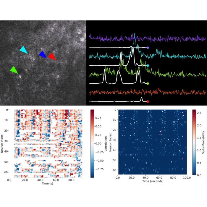
Throughout this course, we have developed a comprehensive workflow for transforming raw calcium imaging data into interpretable representations of neuronal and network dynamics. Our approach has covered all key stages: extracting calcium traces, inferring spike probabilities, and analyzing population-level activity to understand the functional organization of neural circuits.
Revisiting our initial guiding questions, we can now provide the following answers:
How can meaningful neuronal signals be extracted from large-scale calcium imaging data?
Meaningful signals are extracted by identifying and segmenting individual neurons within raw imaging data, then quantifying their fluorescence intensity over time to generate calcium traces. Careful pre-processing and denoising steps are essential to reduce artefacts and isolate the neuronal signals from background and measurement noise. Toolboxes such as CaImAn facilitate this process by providing efficient algorithms for motion correction, segmentation, and trace extraction.
What are the principles and limitations of inferring spike activity from calcium fluorescence traces?
Spike inference transforms indirect, temporally blurred calcium signals into estimates of underlying neuronal spiking, typically using biophysical models or, more recently, machine learning approaches. Limitations include:
- Temporal delay and smoothing
- Calcium indicators respond more slowly than the underlying action potentials, introducing a delay and temporal blur to the recorded signal. Fast spike trains are especially difficult to resolve.
- Nonlinearity and saturation
- The fluorescence response is often not linearly proportional to the number of spikes, especially at high firing rates. Saturation of the indicator can mask the precise number of spikes.
- Variability in indicator kinetics
- Different calcium indicators, and even the same indicator in different cellular environments, may have varying rise and decay times, affecting the temporal fidelity of spike inference.
- Noise and measurement artifacts
- Measurement noise, motion artifacts, and fluctuations in background fluorescence can produce changes in the trace that are indistinguishable from real neuronal events, complicating reliable spike detection.
- Lack of one-to-one correspondence between fluorescence events and single spikes
- Calcium imaging does not provide a direct mapping between each spike and each observed fluorescence event:
- Missed spikes: Some action potentials may not result in detectable changes in fluorescence, especially if the indicator is not sensitive enough or noise is high.
- Merged events: Multiple spikes in rapid succession can lead to overlapping or summed calcium signals, making it impossible to distinguish the exact number or timing of spikes.
- Non-spike events: Occasionally, fluctuations in the trace may not be caused by action potentials at all, but rather by noise, movement, or other cellular processes.
While modern spike inference algorithms can robustly estimate neuronal activity patterns from calcium traces, the process is inherently limited by the indirect, noisy, and temporally filtered nature of the measurement. Results should always be interpreted with these constraints in mind, and, where possible, validated against ground truth data.
Which analytical methods reveal patterns and structure in the activity of neural populations?
Methods such as population activity visualization (e.g., heatmaps), temporal binning, dimensionality reduction (PCA, t-SNE, UMAP), clustering, and correlation-based analyses allow the detection of patterns, assemblies, and functional structure within the ensemble activity. Sorting neurons by their relationship to behavioral variables or population-level signals further enhances interpretability.
How do collective dynamics and functional assemblies emerge from the activity of individual neurons?
Collective dynamics arise when groups of neurons exhibit coordinated or structured patterns of activity, often reflecting underlying network connectivity or engagement with behavioral or experimental conditions. Functional assemblies can be detected as clusters or sequences of co-active neurons, often revealed through population analysis and correlation measures, and reflect the circuit-level computation and information processing performed by the neural network.
Conclusion
In summary, you have learned how to extract and interpret neuronal signals from complex imaging data, apply principled spike inference methods, and analyze neural population activity to uncover patterns, assemblies, and functional organization. These analytical skills provide a solid foundation for exploring brain circuits, relating neural activity to behavior, and conducting further investigations in systems neuroscience.
