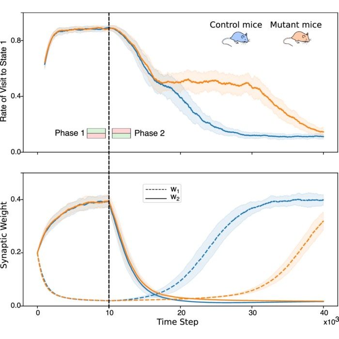
Virchow’s Cellularpathologie: A foundational work in the history of medicine and neuroscience
After that talk by Kettenmann, I could not quite let go of the remaining historical connections that he had mentioned. One of them was the fact that Rudolf Virchow, the founder of cellular pathology, had introduced the distinction between neurons and glial cells in the mid 1850s. And this distinction, as Kettenmann emphasized, was crucial for the later development of neuroscience, because it provided the conceptual framework for understanding the cellular composition of the nervous system.
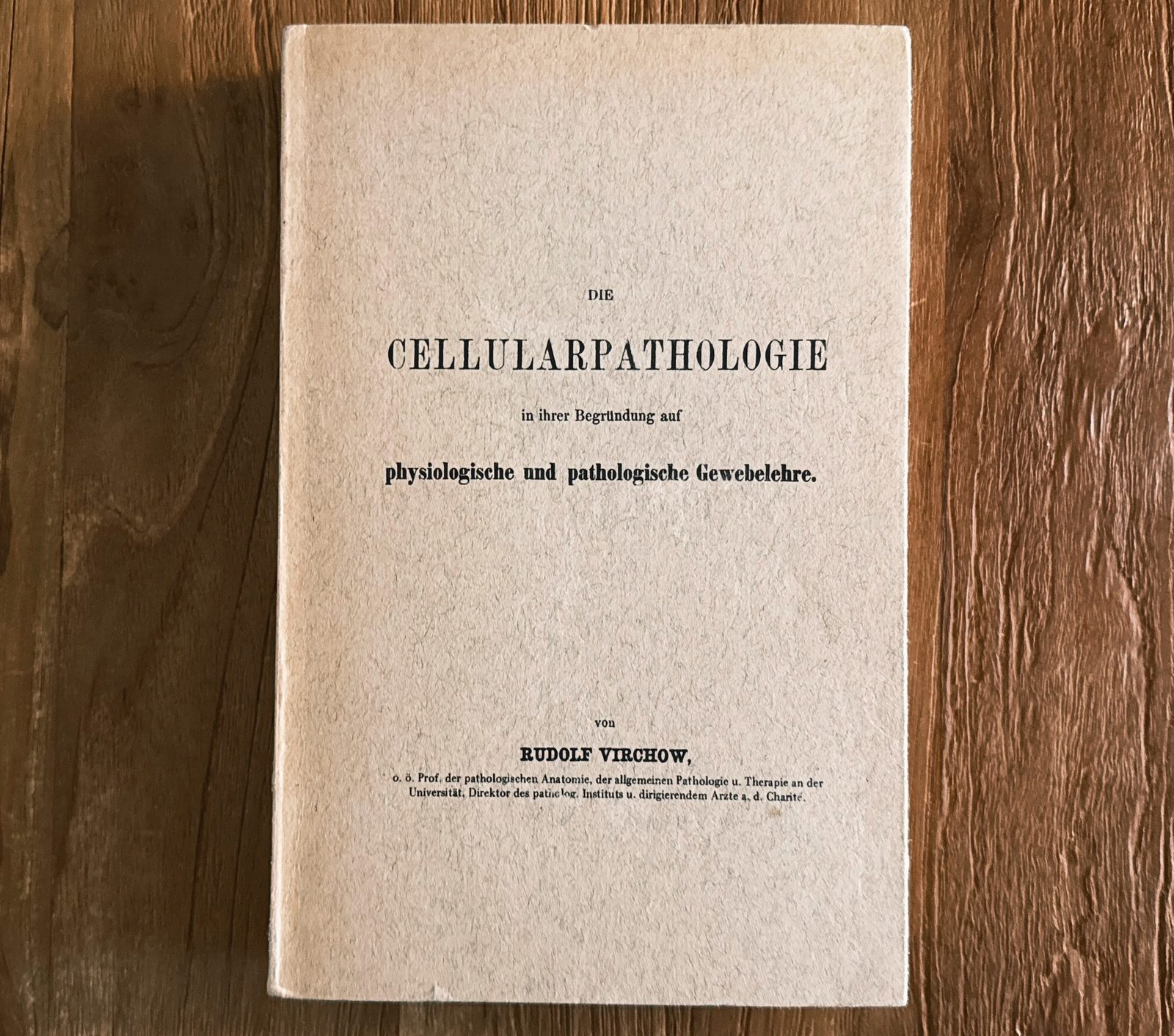
The cover of my personal copy of Virchow’s Cellularpathologie, which I ordered from a second-hand book store. The book is in German, but there may be English translations available as well.
I could not resist buying a physical copy of Virchow’s Cellularpathologie. A used reproduction from 1956, nothing special. But it felt oddly appropriate. As someone still relatively new to neuroscience, I did not know this work before. However, as a side note, I had already encountered Virchow’s name almost on a daily basis: In the city where I grew up, I lived in a neighbourhood where almost all streets were named after German scientists. One of them was Virchowstraße1 (Virchow Street), which I passed by every day on my way to school. Of course, I knew what Virchow was famous for, but I had never read any of his work.
Zoom onto the cover of Virchow’s Cellularpathologie (my personal copy).
I did not read the entire book, since it is quite long and dense, and, from a physicist’s perspective, several chapters are out of my depth. However, since it was written in German, it was of course no problem to understand the text. I found it fascinating how structured and precise this early scientific work is. It felt almost like reading a modern scientific book. Of course, the figures were not made with modern software, as we would do today. Instead, woodcut illustrations were used, which achieve such a high level of detail and clarity that they even surpass some modern illustrations in my view. The book is organized into 20 lectures, each focused on a specific aspect, and the arguments are built step by step, with careful attention to evidence and logical structure.
In this post, I want to share some of the insights I gained from engaging with Virchow’s Cellularpathologie. I will not attempt to summarize the entire book, but rather focus on some key themes and implications that I found interesting.
Virchow and the conceptual shift in medicine
Rudolf Virchow (1821–1902) is often regarded as one of the most influential figures in the history of medicine. He was a physician, pathologist, anthropologist, politician, and social reformer, and his work fundamentally changed how disease was understood in the 19th century. Today, Virchow is primarily remembered as the founder of cellular pathology, but his scientific influence extended much further. He contributed to histology, pathology, epidemiology, and public health, argued strongly for the social dimension of medicine, and introduced or popularized concepts and terms that remain central to medicine today, including thrombosis, embolism, leukemia, and neuroglia.
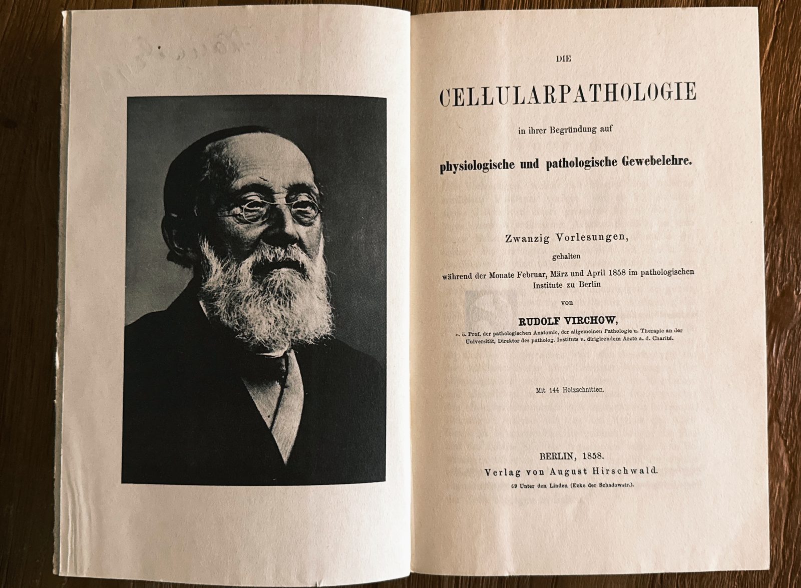
First pages of Virchow’s Cellularpathologie, with a photograph of Virchow himself on the left. Source: Reproduced from Rudolf Virchow, Die Cellularpathologie in ihrer Begründung auf physiologische und pathologische Gewebelehre, 1858. Photograph of the printed figure taken by myself. Original work in the public domain.
His most influential work, however, was undoubtedly Cellularpathologie, first published in 1858. The book laid the conceptual foundation for modern pathology and had profound implications for how disease itself was understood.
Virchow’s central idea is often summarized in a single statement:
Omnis cellula e cellula
Every cell arises from another cell.
At first glance, this sounds almost trivial from a modern perspective. However, in the mid 19th century, this statement directly contradicted older ideas about spontaneous generation and the formation of tissues from amorphous substances. It established continuity at the cellular level as the fundamental principle of life. More importantly, Virchow extended this principle from physiology to pathology. Disease, in his view, is not a disturbance of abstract bodily “fluids” or humors such as blood, phlegm, black bile, and yellow bile, as in ancient medicine, but a disturbance of cells. This is the conceptual core of cellular pathology.
This insight has profound implications: If every physiological process is rooted in cellular processes, then every pathological process must also be reducible to changes in cellular structure, function, or proliferation. In modern terms, this marks a shift from descriptive medicine at the level of organs and symptoms toward a mechanistic and microscopic understanding of disease.
The lectures
Virchow’s Cellularpathologie is not a monolithic treatise in the traditional sense. It is a collection of 20 lectures, delivered in early 1858 at the Pathological Institute in Berlin and later transcribed and edited for publication. This also explains the style of the work: It is didactic, structured around demonstrations, and closely tied to visual material such as microscopic preparations and woodcut illustrations.
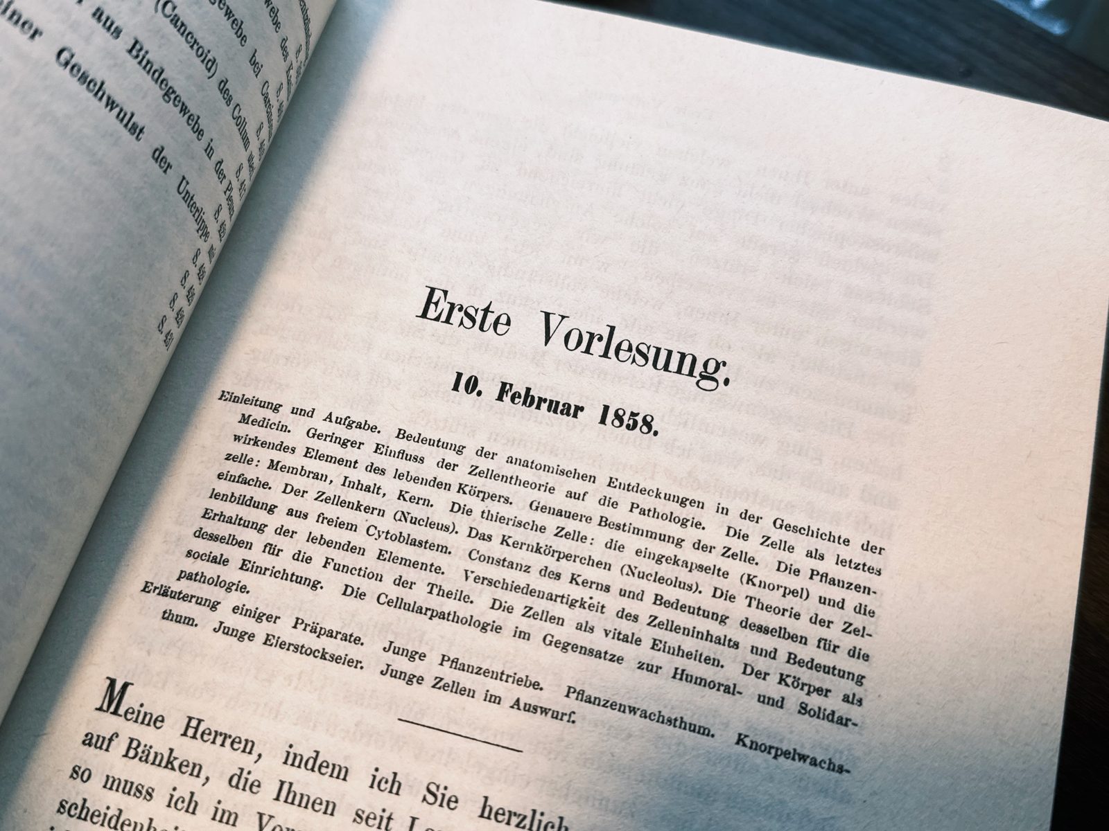
Cover of the first lecture of Virchow’s Cellularpathologie. Source: Woodcut illustration reproduced from Rudolf Virchow, Die Cellularpathologie in ihrer Begründung auf physiologische und pathologische Gewebelehre, 1858. Photograph of the printed figure taken by myself. Original work in the public domain.
The work begins with a systematic overview of cell theory, integrating botanical and zoological observations. The inclusion of plant cells alongside animal tissues reflects the universality of the cellular principle. The same structural logic applies across living systems, whether in plant tissue, cartilage, liver, or nervous tissue, as illustrated in the early figures of the book.
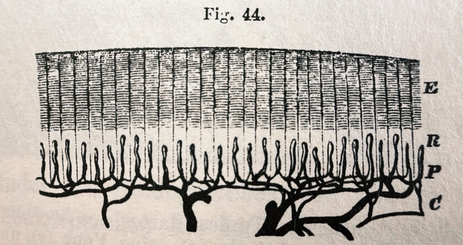
Woodcut illustration of an injection preparation of the skin, vertical cut. E: Epidermis, R: Rete Malpighii, P: Papilla with its up and down projecting vessels, C: Cutis. Source: Woodcut illustration (Fig. 44) reproduced from Rudolf Virchow, Die Cellularpathologie in ihrer Begründung auf physiologische und pathologische Gewebelehre, 1858. Photograph of the printed figure taken by myself. Original work in the public domain.
From this foundation, Virchow gradually develops the framework of cellular pathology in detail. Much of the book can be understood as a systematic attempt to establish the cell as the central explanatory unit of medicine.
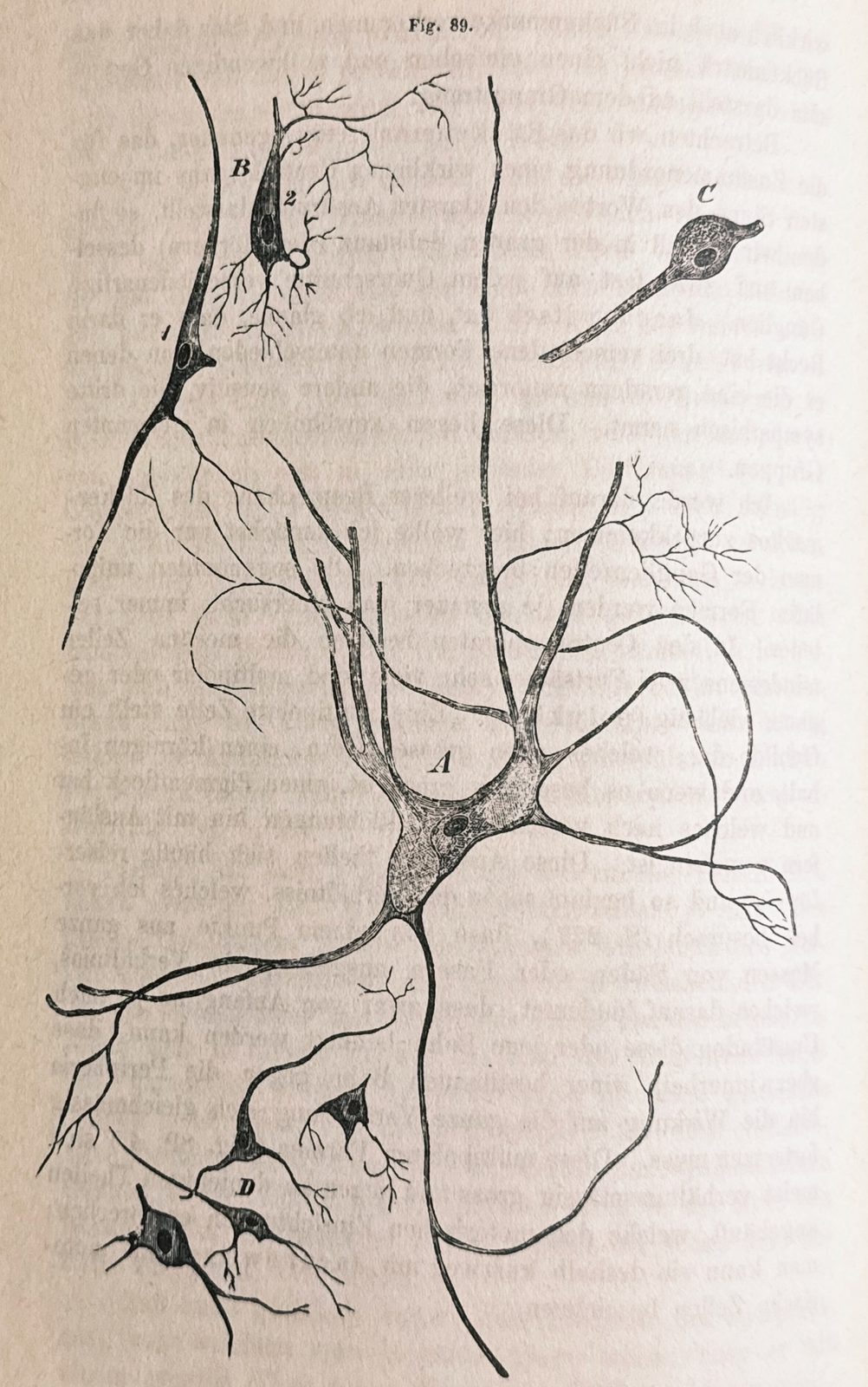
Woodcut illustration of Ganglion cells from the central organs: A, B, C from the spinal cord, based on preparations by Mr. Gerlach; D from the cerebral cortex. A: Large, multi-dendritic (multipolar, polyclonal) cells from the anterior horns (motor cells). B: Smaller cells with 3 larger processes from the posterior horns (sensory cells). C: Two-branched (bipolar, diclonal), more rounded cells from the vicinity of the posterior commissure (sympathetic cells). Magnification: 300. Source: Woodcut illustration (Fig. 89) reproduced from Rudolf Virchow, Die Cellularpathologie in ihrer Begründung auf physiologische und pathologische Gewebelehre, 1858. Photograph of the printed figure taken by myself. Original work in the public domain.
In the lectures, Virchow explicitly argues against older humoral and organ-centered views of disease and instead localizes pathological processes at the cellular level. However, what is particularly interesting here is how concretely he develops this argument. Rather than presenting a purely abstract theory, he continuously moves between microscopic observations, tissue morphology, pathological examples, and conceptual interpretation.
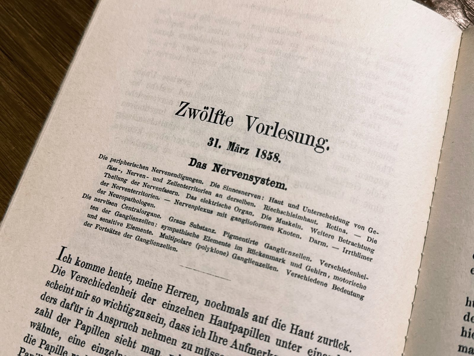
Cover of the 12th lecture of Virchow’s Cellularpathologie, which is focused on the nervous system. Source: Woodcut illustration reproduced from Rudolf Virchow, Die Cellularpathologie in ihrer Begründung auf physiologische und pathologische Gewebelehre, 1858. Photograph of the printed figure taken by myself. Original work in the public domain.
Similarly, Virchow repeatedly emphasizes that pathological processes are not fundamentally separate from physiological ones. Disease is not treated as something metaphysically distinct from normal life processes, but as an alteration, dysregulation, or exaggeration of normal cellular activity. In his lectures, he establishes this view not only philosophically, but through numerous examples involving growth, degeneration, inflammation, and tissue remodeling. The detailed discussion of inflammation is especially interesting here: Inflammation is no longer described merely through externally visible symptoms such as heat, redness, and swelling, but as a sequence of cellular events involving the accumulation, transformation, and activity of specific cells. Likewise, tumors are interpreted not as foreign entities or parasitic growths, but as the result of abnormal cellular proliferation originating from the body’s own tissues.
And Virchow reinforces his arguments with an, at that time, extensive use of microscopic images, which range from plant cells to cartilage, liver cells, capillaries, ganglion cells, and pathological tissues:
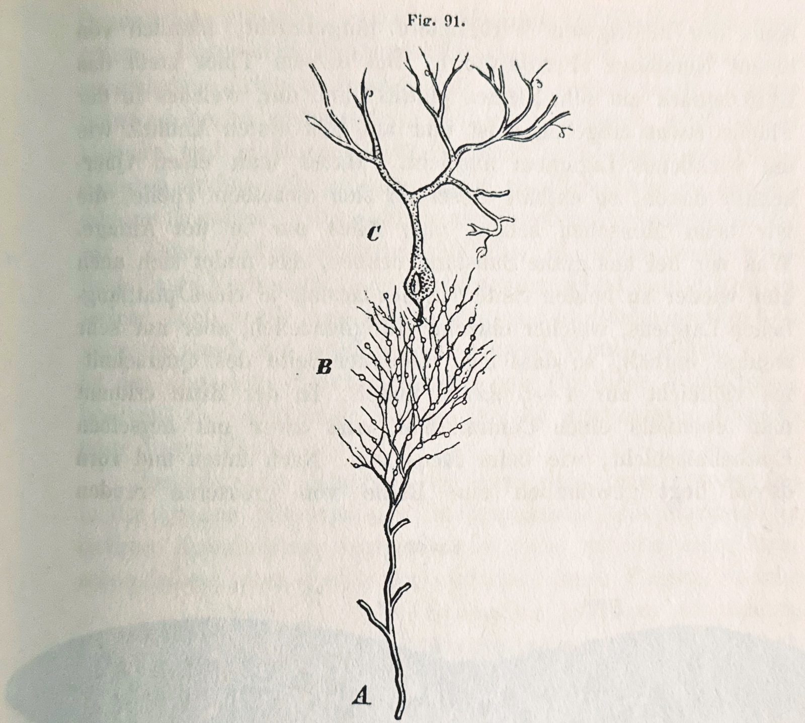
Schematic representation of nerve fiber patterns in the cerebellar cortex according to Gerlach. (Microscopic Studies, Plate I, Fig. 3.) A: white matter; B, C: gray matter; B: granular layer; C: cell layer. Source: Woodcut illustration (Fig. 91) reproduced from Rudolf Virchow, Die Cellularpathologie in ihrer Begründung auf physiologische und pathologische Gewebelehre, 1858. Photograph of the printed figure taken by myself. Original work in the public domain.
Another important aspect is Virchow’s insistence on continuity. There is no abrupt transition from health to disease at the level of fundamental principles. Instead, there exists a continuous spectrum of cellular states and transformations. This idea feels remarkably modern, since many diseases today are understood not as entirely separate biological phenomena, but as dysregulated versions of normal cellular processes.
Finally, and in direct connection to our earlier discussion of Helmholtz, Virchow introduced the concept of neuroglia as a distinct cellular component of nervous tissue. This distinction between neurons and glial cells provided an important conceptual framework for later neuroscience and fundamentally changed how the nervous system was understood at the cellular level. What Virchow recognized was that the nervous system could not be reduced to nerve fibers and nerve cells alone. It also contained another cellular and structural component, which he interpreted as a kind of connective or supporting tissue of the nervous system. The term neuroglia itself expresses this idea: A “nerve glue”, a tissue that holds the nervous elements together:
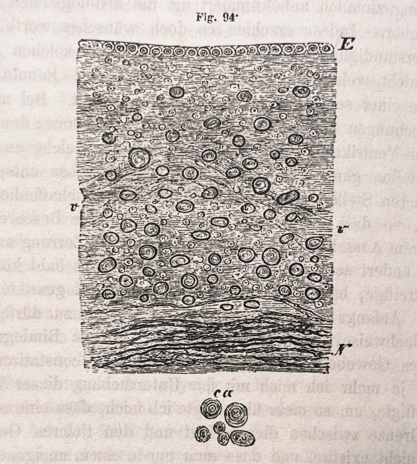
Woodcut illustration of the ependyma of the ventricles and neuroglia from the floor of the fourth ventricle. E: epithelium, N: nerve fibers. In between lies the free portion of the neuroglia, containing numerous connective tissue cells and nuclei; at v is a blood vessel; elsewhere, numerous corpora amylacea are visible, which are shown in isolation at ca. Magnification: 300. Source: Woodcut illustration (Fig. 94) reproduced from Rudolf Virchow, Die Cellularpathologie in ihrer Begründung auf physiologische und pathologische Gewebelehre, 1858. Photograph of the printed figure taken by myself. Original work in the public domain.
From a modern perspective, this interpretation was still limited, because glial cells are no longer understood as merely passive support elements. Astrocytes, oligodendrocytes, microglia, and other glial cell types are now known to participate actively in homeostasis, metabolism, myelination, immune defense, synaptic regulation, and disease processes. But Virchow’s conceptual step was nevertheless crucial: He separated the nervous system into more than one cellular category and thereby opened the possibility of asking what these non-neuronal cells are, where they come from, and what they do.
Conceptual implications
What I take away from researching and reading Virchow’s Cellularpathologie is that it represents a major conceptual shift in the history of medicine. It is not just a collection of observations or a description of pathological phenomena. It is an attempt to reorganize the entire field of medicine around the cell as the fundamental unit of life and disease. Virchow constructs a coherent explanatory framework that connects anatomy, physiology, pathology, microscopy, and tissue structure into a unified view of disease. In this sense, the book can be regarded as on of the scientifically most important works in the history of medicine, and it laid the groundwork for many subsequent developments in pathology, histology, and neuroscience.
Several aspects of Virchow’s reasoning still feel surprisingly familiar, at least from what I can recognize as a non-medical reader. The idea that disease emerges from altered cellular states, that pathological and physiological processes are deeply connected, and that microscopic structures can provide mechanistic explanations for macroscopic symptoms remains central to biomedical science today.
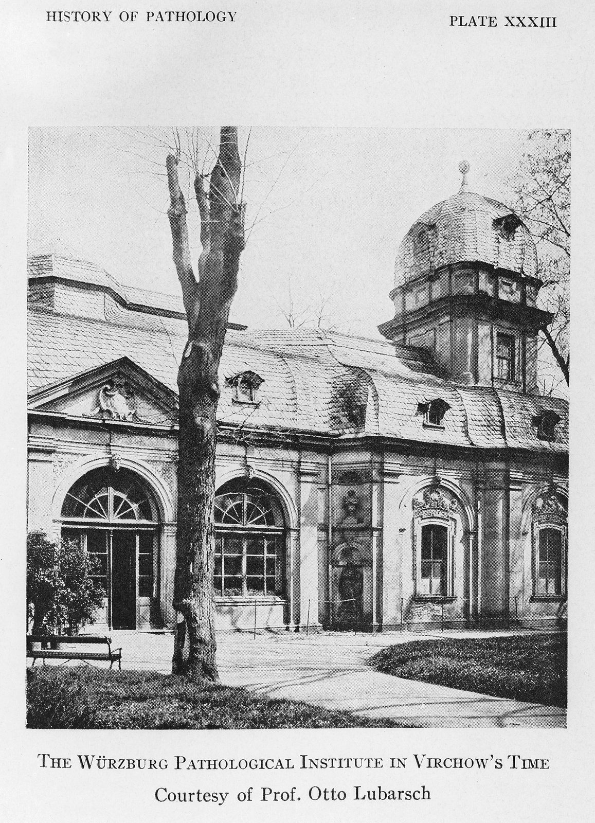
The garden pavilion of the Juliusspital in Würzburg housed Virchow’s Institute of Pathology until 1853. Source: Wikimedia Commonsꜛ (license: CC BY-SA 4.0).
Engaging with Virchow after reading Helmholtz’s dissertation also highlights an interesting historical contrast. Helmholtz’s dissertation is to some extent a bit more observational and comparative. Virchow, by contrast, is much more focused on conceptual integration and theoretical organization. He builds a framework in which such structures acquire general pathological meaning. In this sense, the two works together provide an interesting glimpse into the emergence of modern life sciences during the 19th century, which could, in a very simplified way, be seen as: One focused on observation and comparative anatomy, the other on conceptual unification and medical interpretation.
This little excursion into the history of science was quite refreshing. Ideas we now take for granted, not only in neuroscience and medicine, must have been revolutionary at that time and had to be established through careful argumentation and evidence, which is not so different from today. I also enjoyed reading about the biographies of 19th and early 20th century2 scientists, who were often polymaths3 and engaged in multiple fields of science, medicine, and even politics. This again reminds me that the boundaries between disciplines were much more fluid at that time, and that scientific progress often involved contributions from individuals with broad interests and expertise. An attitude that I think is worth remembering today.
References and further reading
- Rudolf Virchow, Die Cellularpathologie in ihrer Begründung auf physiologische und pathologische Gewebelehre: Zwanzig Vorlesungen, gehalten während der Monate Februar, März und April 1858 im pathologischen Institute zu Berlin, 1858, Verlag von August Hirschwald, Berlin; online available for free at MDZ Digitale Sammlungenꜛ or on archive.orgꜛ (original work in the public domain)
- Julia Heideklang, H.-J. Pflüger, Helmut Kettenmann, De fabrica systematis nervosi evertebratorum. Die kommentierte Dissertation von Hermann Helmholtz, 2021, Wbg Academic, ISBN: 9783534400942, online PDFꜛ, Websiteꜛ
Footnotes
-
And guess what, Helmholtzstraße (Helmholtz Street) was only a few streets away, and I passed by it almost every day as well. So, in a way, perhaps it was inevitable that I would eventually engage with the natural sciences. There were many other streets named after scientists, too, but no worries, I will not turn this blog into a tour through the scientific street names of my childhood town. ↩
-
See, e.g., our post on Richard Feynman’s problem solving approach. ↩
-
See, e.g., this Mastodon postꜛ. ↩

comments